Photoacoustic microscopy (PAM) is a relatively new imaging technique that uses laser light to induce ultrasonic vibrations in tissue. These ultrasonic vibrations, along with a computer that processes them, can then be used to create an image of the structures of the tissue in much the same way ultrasound imaging works.
In the last few years, Lihong Wang, Caltech’s Bren Professor of Medical Engineering and Electrical Engineering, has developed PAM technologies that can image changing blood flow in the brain, detect cancerous tissue, and even identify individual cancer cells.
However, one limitation of high-resolution (i.e., optical-resolution) PAM has been its narrow depth of field, meaning that it can only focus on a thin layer (approximately 30 micrometers, or about the length of one skin cell, with one to two micrometers of resolution) of tissue at a time. To see something above or below the plane that the device is viewing, it needs to refocus above or below that plane. For comparison, imagine a person putting on reading glasses to do a crossword puzzle.
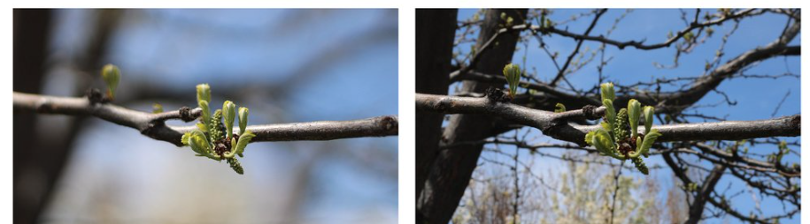
Depth of field: At left, an image of a tree branch taken with a shallow depth of field. Only the foreground is in focus. At right, an image of the same branch taken with a deep depth of field. Both the foreground and background are in focus.
Credit: NightWolf1223/Wikimedia Commons
In a new paper in the journal Nature Photonics, Wang and his research team show how they developed a new variant of PAM called needle-shaped beam photoacoustic microscopy, or NB-PAM, which that has a depth of field nearly 14 times greater than what was achievable before. This means NB-PAM can create 3-D imagery of samples without refocusing and better image samples with uneven surfaces.
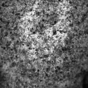
An image of mouse liver tissue taken with traditional photoacoustic microscopy (left) compared to an image of mouse liver tissue taken with needle-shaped beam photoacoustic microscopy (right). In the image at the right, structures are in focus over a greater range of depth.
Credit: Caltech
“Some applications, such as studying tissue samples without needing to use a microscope slide, require imaging of uneven surfaces at high spatial resolution,” says Rui Cao, lead author and postdoctoral scholar research associate in medical engineering. “Conventional PAM grapples with the trade-off between resolution and depth of field, which has been overcome by our new technology.”
NB-PAM improves its depth of field over its related PAM technologies by using a beam of laser light that is longer and thinner, hence “needle shaped.” This change in the optical characteristics of the beam avoids some of the drawbacks associated with other attempts to increase the depth of field of PAM technology, such as working more slowly, or requiring more computer processing power.
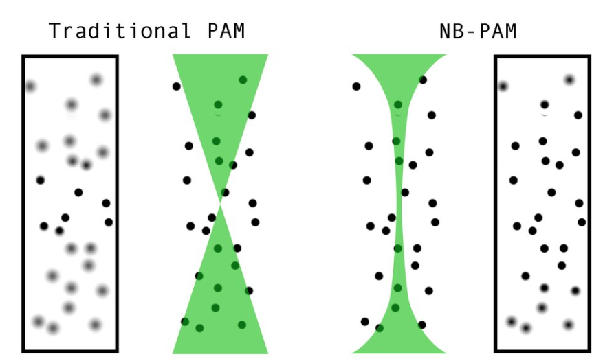
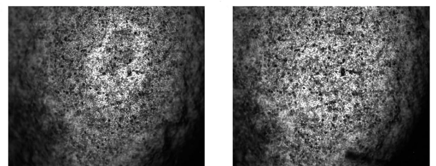
Traditional photoacoustic microscopy (PAM) (left) compared to needle-shaped photoacoustic microscopy (NB-PAM) (right). In traditional PAM, only objects near the focal point of the laser are imaged sharply. In NB-PAM, the longer, narrower beam allows objects over a greater range of depth to be clearly imaged.
Credit: Caltech
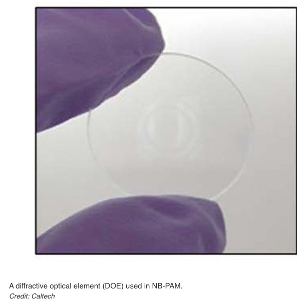
This needle-shaped beam is created using a specialized item known as a diffractive optical element (DOE). To the casual observer, a DOE looks like a tiny sheet of glass, but it is actually a piece of fused silica with precise patterns engraved on its surface. Those patterns reshape the beam of light that is used for imaging, so that it no longer focuses to a sharp point along the propagation axis but is instead drawn out into a long thin neck. Consequently, it is able to clearly image objects over a greater range of depths.
That greater depth of field was demonstrated by the researchers in two ways: imaging fresh organ samples using an ultraviolet laser and imaging in vivo mouse brain vasculature using a blue laser.
“This technology provides new opportunities for studying tissue samples during surgery, which would allow complete removal of cancer cells and maximal preservation of normal ones,” says Wang, who is also the Andrew and Peggy Cherng Medical Engineering Leadership Chair; Executive Officer for Medical Engineering. “Translation into the operating room is a natural future avenue of research.”

Rui Cao, lead author and postdoctoral scholar research associate in medical engineering
Credit: Rui Cao
The paper describing NB-PAM, titled “Optical-resolution photoacoustic microscopy with a needle-shaped beam,” appears in the December 1 issue of Nature Photonics. Co-authors are Lei Li (PhD ’19), Yide Zhang, and Samuel Davis, all postdoctoral scholar research associates in medical engineering; medical engineering graduate student Yilin Luo; Jingjing Zhao and Adam de la Zerda of Stanford University; Lin Du of University of Pennsylvania; and Qifa Zhou and Laiming Jiang of USC.
Funding for the research was provided by the National Institutes of Health.
WRITTEN BY
Emily Velasco
CONTACT
Emily Velasco
(626) 372‑0067
evelasco@caltech.edu
出典:
https://www.caltech.edu/about/news/seeing-more-with-a-needle-shaped-laser
In case of addition or removal of the article, please contact us as below:
info@symphotony.com
この情報へのアクセスはメンバーに限定されています。ログインしてください。メンバー登録は下記リンクをクリックしてください。