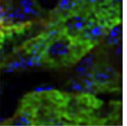
Researchers could rapidly obtain high-resolution images of blood vessels and neurons within the brain.
Anne Trafton | MIT News Office
Publication Date:July 7, 2021
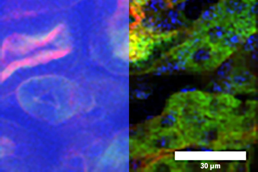
To create high-resolution, 3D images of tissues such as the brain, researchers often use two-photon microscopy, which involves aiming a high-intensity laser at the specimen to induce fluorescence excitation. However, scanning deep within the brain can be difficult because light scatters off of tissues as it goes deeper, making images blurry.
Two-photon imaging is also time-consuming, as it usually requires scanning individual pixels one at a time. A team of MIT and Harvard University researchers has now developed a modified version of two-photon imaging that can image deeper within tissue and perform the imaging much more quickly than what was previously possible.
この情報へのアクセスはメンバーに限定されています。ログインしてください。メンバー登録は下記リンクをクリックしてください。